. 2025 Jun 6;15(1):19969.
doi: 10.1038/s41598-025-02424-x.

Femoral strength after cephalomedullary nail removal can be predicted preoperatively using CT based FE models

Affiliations

Femoral strength after cephalomedullary nail removal can be predicted preoperatively using CT based FE models

Alexander Synek et al. Sci Rep. .

Abstract

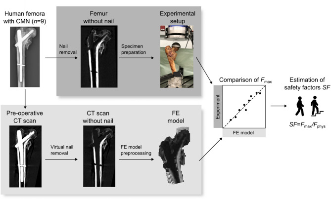
Removals of cephalomedullary nails (CMNs) after healed pertrochanteric femur fractures are sometimes requested by patients or medically indicated due to pain or screw cut-out. However, CMN removal carries a high risk of secondary femoral neck fracture, even in the absence of trauma. Consequently, decisions on nail removal and establishing a safe post-operative loading regimen can be challenging. This study investigated if finite element (FE) models can pre-operatively predict femoral strength after CMN removal to support these clinical decisions. Nine proximal femora of body donors who were treated with a CMN during their lifetime were included. Computed tomography (CT) scans were acquired with the CMN still in place, followed by virtual implant removal using image processing. Based on this scan, non-linear voxel-based FE models were created and femoral strength was predicted for a one-legged stance configuration. For validation, the CMNs were physically removed and femoral strength was assessed in a material testing machine. The FE models predicted the femoral strength accurately relative to the experiments (R2 = 0.94, CCC = 0.97). In conclusion, CT-based FE models demonstrate potential to predict femoral strength after CMN removal pre-operatively. This could help patients and clinicians to make an informed decision on implant removal and permissible post-operative weight-bearing.

Keywords: Cephalomedullary nail; Femur; Finite element; Fracture risk; Implant removal.


Conflict of interest statement

Declarations. Competing interests: Dieter H. Pahr is CEO of Dr. Pahr Ingenieurs e.U., which develops and distributes Medtool. All other authors declare no competing interests.

Figures

Fig. 1
Fig. 1
Outline of the study. Nine human proximal femora with implanted cephalomedullary nails (CMN) were used. Femoral strength (Fmax) after nail removal in one-legged stance configuration was compared between the experimental measurement and the pre-operative FE model prediction. Safety factors (SF) were estimated for clinical interpretation by dividing Fmax with the estimated physiological peak force (Fphys) during various activities.
Fig. 2
Fig. 2
Anterior-posterior radiographs of the nine proximal femora used in this study.
Fig. 3
Fig. 3
Pre-operative CT scan before and after virtual nail removal for one representative specimen. A grey value profile along vector d, and a cross section at the distal locking screw are shown. At the distal locking screw, blooming and streak artefacts were visible.
Fig. 4
Fig. 4
Details of one representative FE model. The model was cropped to 60% of the specimen height L and element-specific material properties were assigned based on the bone volume fraction (ρ). A reference node was inserted at the centre of a sphere fitted to the proximal embedding. All nodes at the distal end of the femur were constrained and a displacement u was imposed on the reference node R.
Fig. 5
Fig. 5
Comparison of experimentally measured and FE-predicted femoral strength (Fmax). Coefficient of determination (R2), concordance correlation coefficient (CCC) and the line equation of the linear regression are shown. Labels of individual specimens are displayed next to the data points.
Fig. 6
Fig. 6
Comparison of the estimated fracture locations observed in the experiments to the damaged regions in the FE models. Fracture locations of the experiments were estimated based on CT scans taken after testing, visual inspection of the specimens, and video recordings. The damaged regions of the FE models are displayed based on a frontal cross section at the last time increment of the simulation.
Fig. 7
Fig. 7
Estimated safety factors SF75 for all nine femora and different daily activities. Safety factors were computed by dividing the predicted femoral strength of the FE models (Fmax) with the averaged in vivo peak hip joint load of subjects scaled to 75 kg body weight (taken from Bergmann et al.). Femora and activities which reached a safety factor SF100 ≥ 1 are highlighted with * (i.e. using in vivo hip joint loads of the subject with highest relative loads and scaled to 100 kg body weight from Bergmann et al.).

References

    1. Nherera, L., Trueman, P., Horner, A., Watson, T. & Johnstone, A. J. Comparison of a twin interlocking derotation and compression screw cephalomedullary nail (InterTAN) with a single screw derotation cephalomedullary nail (proximal femoral nail antirotation): A systematic review and meta-analysis for intertrochanteric fractures. J. Orthop. Surg. Res.13, 1–10 (2018). - DOI - PMC - PubMed
    1. Ng, M. et al. No difference between lag screw and helical blade for cephalomedullary nail cut-out a systematic review and meta-analysis. Eur. J. Orthop. Surg. Traumatol.32, 1617–1625 (2022). - DOI - PubMed
    1. Kukla, C., Gaebler, C., Mousavi, M., Vecsei, V. & Heinz, T. Indications for implant removal in healed proximal femoral fractures. Acta Chir. Austriaca32, 196–198 (2000). - DOI
    1. Barquet, A., Giannoudis, P. V. & Gelink, A. Femoral neck fractures after removal of hardware in healed trochanteric fractures. Int. J. Care Injury48, 2619–2624 (2017). - DOI - PubMed
    1. Yang, J. H. et al. The Analysis of Biomechanical Properties of Proximal Femur after Implant Removal. Appl. Bionics Biomech. (2016). - PMC - PubMed